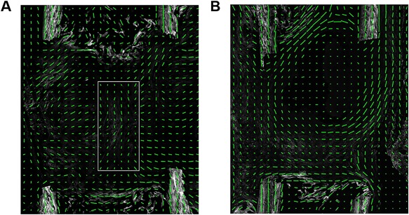
Figure 6.

Matrix alignment. (A) Flexible group at day 14. Fiber orientation in the middle of the defect (white box) runs in parallel to the bone axis (cf, Supplemental Fig. S9A). (B) Semi‐rigid group at day 14. Fiber orientation at the margins of the fracture site runs in parallel to the bone axis and perpendicular fibers seal the medullary canal (cf, Supplemental Fig. S9B). (A, B) A green line in each sub‐ROI indicates the direction of primary fibril orientation, and the length of the line represents the degree of anisotropy.
