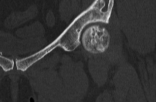
Figure 3.

CT left hip/pelvis coronal view: Irregular mixed lucent and sclerotic lesion in the femoral head correspond to the subchondral lesion found on MRI. This is associated with a 3‐mm cortical breach at the anterosuperior margin of the femoral cortex.
