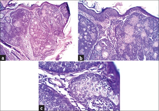
Figure 3.

(a) Ulcerated epidermis focally with an infiltrative growth pattern of tumor in dermis (H and E, ×40) (b) Many undifferentiated cells with sebaceous cells having foamy cytoplasm (H and E, ×100) (c) The undifferentiated cells have round-to-oval pleomorphic nuclei with dispersed chromatin and eosinophilic cytoplasm (H and E, ×400)
