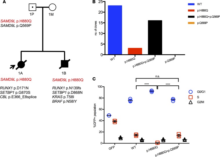
Figure 2. SAMD9L H880Q mutation in MLSM7 family 1.
(A) Pedigree showing the germline SAMD9L mutation (red font) in the father (1P) and patients 1A and 1B, as well as secondary somatic mutations identified in their AML bone marrows (black font). Squares indicate male family members, circles female members. Unaffected individuals are indicated with open symbols, unaffected mutation carriers are denoted with black dots, affected individuals with a clinical history of monosomy 7 are shown with filled symbols, and symbols with a slash denote deceased individuals. Arrow indicates the proband. (B) Sequencing data from the father (1P) showing the number of individual amplicons with normal sequence (left), those with the p.H880Q mutation only (middle), and those with the p.H880Q and p.Q569P substitutions in cis (right). The p.Q569P second-site mutation was only detected in the context of the p.H880Q variant and accounted for over 80% of mutant transcripts. (C) EdU cell cycle results showing that 293T cells expressing SAMD9L p.H880Q exhibit a cell cycle arrest phenotype compared with cells expressing the normal SAMD9L protein. This defect is partially corrected by the second-site p.Q569P substitution detected in 1P. Percentages of cells in the S phase of the cell division cycle were compared using 1-way ANOVAs with repeated measures followed by Tukey’s post hoc multiple-comparisons test; significance was based on α = 0.05 (n = 3). ****P < 0.0001. n.s., not significant. Individual data points, and mean ± SD are shown. Data representative of 3 experiments completed in triplicate.

