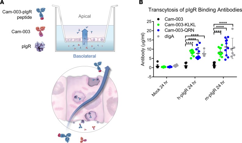
Figure 2. Transcytosis of Cam-003 peptide fusions in pIgR-transfected MDCK cells.
Antibodies were added to the bottom chamber (basolateral) of transwells containing either untransfected, h-pIgR–, or m-pIgR–transfected Madin-Darby canine kidney (MDCK) monolayers (A). At 24 hours, antibodies in the top chamber (apical) were quantified by ELISA. Cam-003 levels were similar in all 3 cell lines, whereas Cam-003–QRN and Cam-003–KLKL, as well as human dimeric IgA, had increased accumulation in the apical chamber (B). Data are representative of at least 2 independent experiments (n = 10 for Cam-003 antibodies, n = 6 for dIgA). Significance was determined using 2-way ANOVA (with Tukey’s post hoc test; ****P < 0.0001).

