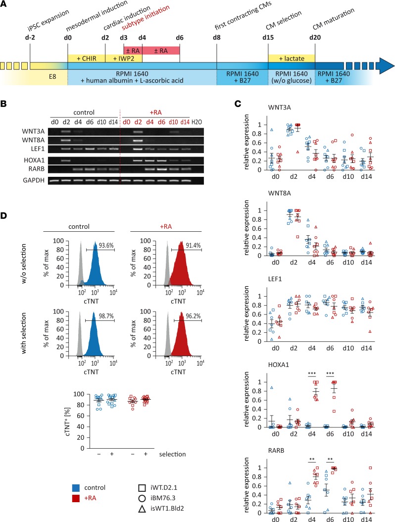
Figure 1. Directed differentiation of iPSCs into atrial or ventricular cardiomyocytes (CMs).
(A) Schematic of the defined differentiation protocols. Small molecules CHIR and IWP2, which modulate canonical WNT signaling, were applied for the induction of iPSCs into CMs. Retinoic acid (RA) was used to induce the atrial subtype specification. Metabolic selection with lactate was applied to achieve a higher purity of iPSC-CMs. (B and C) Expression of genes involved in WNT and RA signaling was assessed by reverse transcriptase PCR analysis in control and RA-treated cells. Shown are representative images (B, iPSC line iBM76.3) and semiquantitative analysis of gene expression (C, n = 2–3 independent differentiation experiments performed for each iPSC line; 3 iPSC lines were used). Results were quantified according to intensity and normalized to GAPDH expression. (D) Flow cytometry analysis of iPSC-CMs with or without lactate selection for cardiac troponin T (cTNT). Top: Representative measurements (iPSC line iBM76.3); gray peaks represent the isotype control. Bottom: Quantitative analysis (n = 11–14 control and n = 9–15 RA-treated independent differentiation experiments from 3 iPSC lines). Data are presented as mean ± SEM. **P < 0.01, ***P < 0.001 by nonparametric Mann-Whitney U test.

