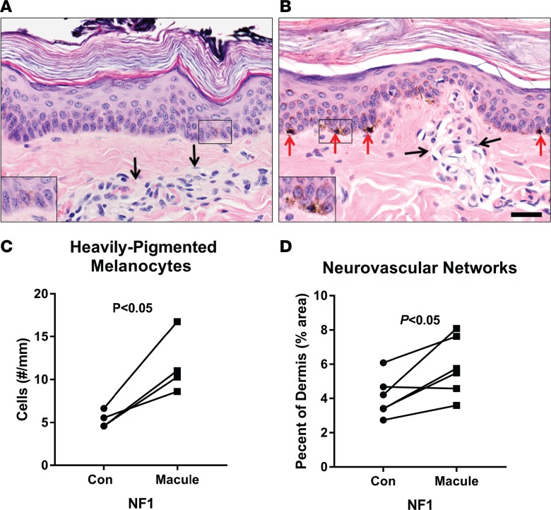
Figure 3. Presence of café au lait macules in newborn NF1 mutant miniswine.
Histopathology of the macules from NF1+/ex42del animals age newborn to <4 months showed increased melanin content within cells of the lower/basal epidermis compared with adjacent control skin from NF1+/ex42del (insets), H&E stains. Skin has scattered pigmented cells along the basal layer (A, black arrows) that are morphologically consistent with melanocytes. Heavily pigmented melanocytes (B, red arrows) were more easily detected in the basal epidermis of macules compared with adjacent skin in NF1+/ex42del miniswine (P < 0.05, paired 2-tailed Student’s t test) (C). Additionally, the dermis of macules compared with adjacent controls skin of NF1+/ex42del miniswine had increased expansion of neurovascular networks (black arrows, A and B) as a percent of dermis area (P < 0.05, paired Student’s t test) (D). n = 4–6; scale bar: 30 μm.

