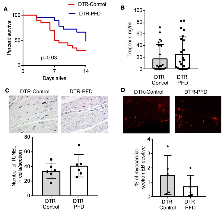
Figure 1. Effect of pirfenidone on mortality and cardiac myocyte cell death after DT treatment.
Mice expressing the diphtheria toxin receptor (DTR) in the myocardium were exposed to diphtheria toxin (DT) and fed either chow enriched with pirfenidone (DTR-PFD) or regular chow (DTR-control). (A) Kaplan-Meier survival curves of DTR control and DTR-PFD mice (n = 20 per group). (B) Serum troponin levels measured at day 4 after DT treatment in DTR-PFD and DTR-control animals (n = 23/group). (C) Cardiac myocyte apoptosis measured at day 4 after treatment with DT. Upper panels are representative histological sections of myocardium from DTR-control and DTR-PFD mice at ×40 magnification. Lower panel summarizes the group data (n = 6 mice/group, 4 sections per animal analyzed). (D) Evans blue (EB) dye uptake at day 4 after DT treatment in DTR-control and DTR-PFD animals; upper panels are representative fluorescence microscopy images at ×10 magnification; lower panel summarizes the group data (n = 5 control; n = 6 mice with pirfenidone; 4 sections per animal analyzed). Bars represent the mean, and error bars represent standard deviation. P values were calculated with the Gehan-Breslow-Wilcoxon method for panel A and with Student’s t test for panels B–D.

