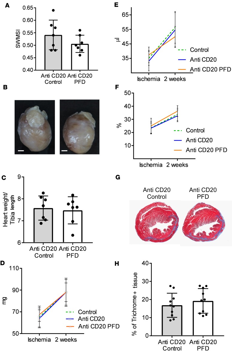
Figure 6. Effect of B cell depletion on pirfenidone cardioprotective effect after I/R injury.
Wild-type mice were B cell depleted via injection of anti-CD20 antibody. Seven days after injection of anti-CD20 antibody, B cell–depleted mice were subjected to 90 minutes of closed-chest ischemia followed by reperfusion (I/R injury). Mice were fed either chow enriched with pirfenidone (anti-CD20 PFD) or regular chow (anti-CD20 control). (A) Area at risk during closed chest-ischemia as determined by the simplified segmental wall motion score index (SWMSI) at time of ischemia. (B) Representative pictures of hearts harvested from anti-CD20–treated mice (left) and anti-CD20 + pirfenidone–treated animals (right). Scale bar: 1 mm. (C) Gravimetric analysis of hearts harvested from pirfenidone-treated animals (anti-CD20 PFD) and untreated controls (anti-CD20 control), n = 7/group. (D–F) Echocardiographic assessment of myocardial function at the time of ischemia and 2 weeks after I/R injury, n = 7/group. Data from non–anti-CD20–treated animals already reported in Figure 3, D–F are shown again for comparison only (control). (D) Left ventricular (LV) mass (LVM) by 2-D echocardiography. (E) LV end-diastolic volume (LVEDV). (F) LV ejection fraction (LVEF). (G) Representative trichrome staining of histological sections of hearts from control animals (left panel) and pirfenidone-treated animals (right panel). Original magnification, ×1.25. (H) Quantitative assessment of the percentage of trichrome-positive staining, 2 sections analyzed per heart, n = 10 sections/group. Bars represent the mean, and error bars represent standard deviation. P values were calculated with Student’s t test.

