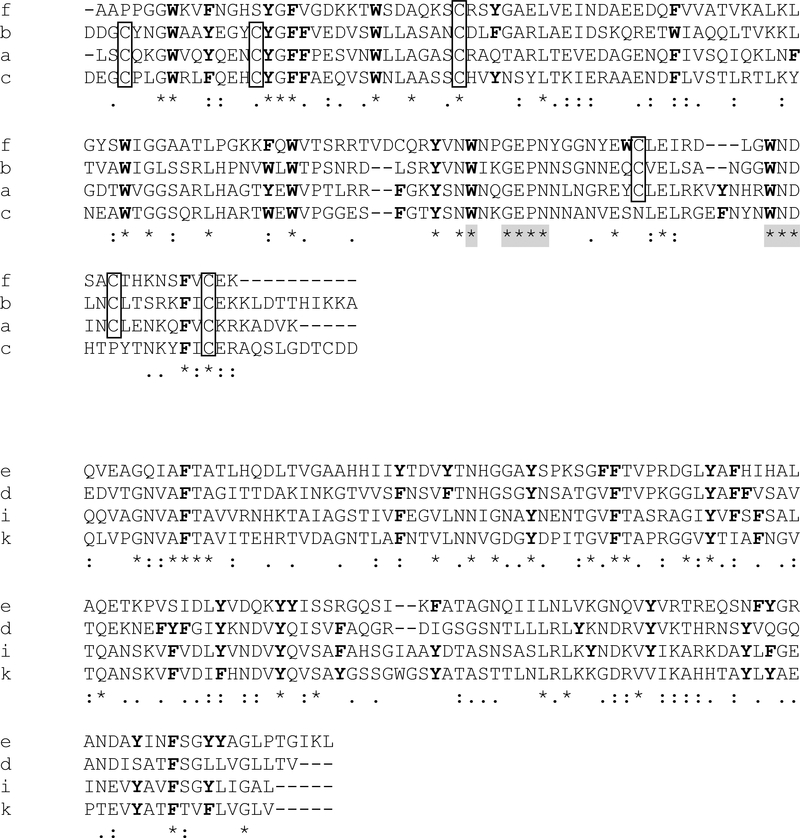
Figure 4.
(a) Clustal alignment of the four C-lectin domain containing proteins. Cysteine residues are boxed. All tryptophan and phenylalanine residues are highlighted in bold, as are tyrosines found at locations where the presence of an aromatic amino acid is conserved. Shaded asterisks indicate matches with consensus calcium and carbohydrate-binding sites. (b) Clustal alignment of C1q-containing proteins, with all tyrosine and phenylalanine residues highlighted in bold. There are no conserved cysteine residues. Asterisks indicate conserved sites, and dots indicate sites that contain amino acids with similar properties. Letters on the left are the letter identifiers appended to Asmp15 to distinguish the different proteins.

