
Fig. 1. H4K16ac is redistributed during normal aging and AD.
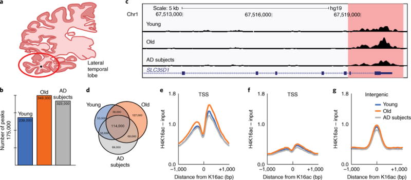
a, Coronal section of human brain indicating the lateral temporal lobe (red circle) used in this study. b, Bar plot of total number of H4K16ac peaks. c, UCSC Genome browser track view of H4K16ac peak at the SLC35D1 gene promoter in Young, Old and AD subjects. d, Venn diagram of peak overlap among Young, Old and AD subjects. e–g, Meta-profile of H4K16ac enrichment at (e) TSSs (±1 kb) of constitutive peaks; (f) TSS (±1 kb) where no peak is detected; and (g) intergenic constitutive peaks (peaks shared across Young, Old and AD subjects).