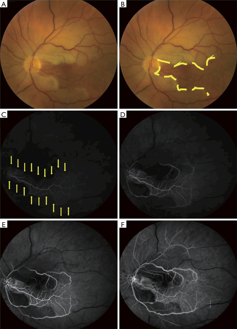
Figure 2.
Left central retinal artery occlusion (CRAO) with sparing of the cilioretinal artery. Color fundus photographs (A,B) and retinal fluorescein angiography (C,D,E,F) in acute left CRAO with cilioretinal artery sparing (same patient as Figure 1). A. Color fundus photograph of the left eye showing a CRAO with cilioretinal artery sparing. The papillomacular bundle is perfused by a patent cilioretinal artery. (B) Same photograph as in (A) outlining the area perfused by the cilioretinal arteries (area contained within the yellow lines). Retinal edema is seen outside of the area perfused by the cilioretinal arteries. (C,D,E,F) Fluorescein angiogram of the left eye taken 21 seconds (C), 24 seconds (D), 28 seconds (E), and 1 minute and 5 seconds (F) after injection of fluorescein dye. There is minimal fluorescent signal in the papillomacular bundle (C, area outlined by yellow arrows) and no appreciable fluorescent signal outside of the papillomacular bundle 21 seconds after fluorescein dye injection. One minute after injection of fluorescein dye (F), there continue to be large vascular segments without fluorescent signal (black segments of retinal vessels) and almost no retinal perfusion outside of the papillomacular bundle.

