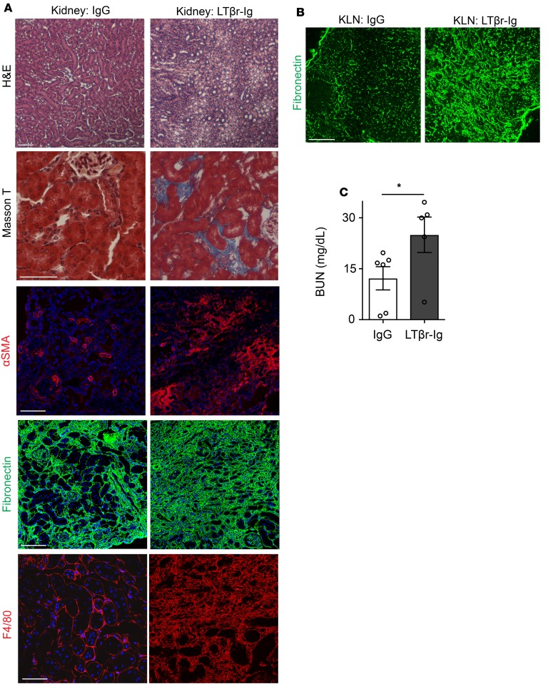
Figure 4. LTβ receptor–immunoglobulin (LTβr-Ig) treatment enhances renal fibrosis following IRI.
(A) Ischemic kidney from mouse treated with LTβr-Ig fails to recover histologically (H&E) 30 days following IRI and shows tissue fibrosis (Masson T), in comparison with the kidney from mouse treated with control IgG. Thirty days following IRI, the ischemic kidney from mouse treated with LTβr-Ig shows increased staining of αSMA, fibronectin, and F4/80, as compared with the kidney from mouse treated with control IgG. Scale bars: 75 μm. (B) Thirty days following IRI, KLN from mouse treated with LTβr-Ig shows marked increase in fibronectin staining, as compared with KLN from mouse treated with control IgG. Scale bar: 200 μm. (C) Thirty days following IRI, BUN is significantly higher in mouse receiving LTβr-Ig, as compared with mouse treated with control IgG (n = 5–6/group, mean ± SEM). *P < 0.05 by Student’s t test.

