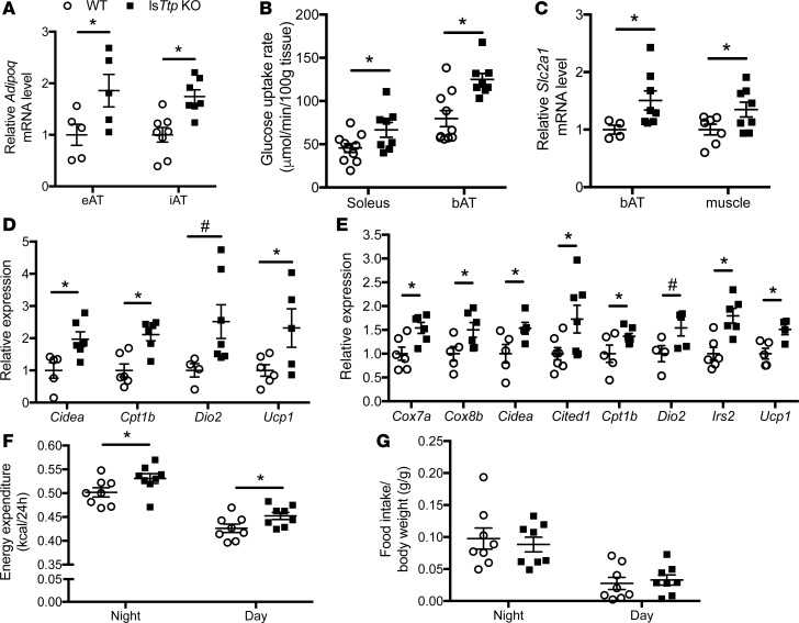
Figure 6. Hepatic deletion of Ttp activates brown and beige adipose tissue.
(A) Adipoq mRNA levels in epididymal adipose tissue (eAT) and inguinal AT (iAT) from WT and lsTtp-KO mice fed HFD (n = 5–8). (B) Soleus muscle and brown adipose tissue (bAT) glucose uptake at the end of hyperinsulinemic-euglycemic clamp studies in WT and lsTtp-KO mice fed HFD (n = 7–10). (C) Slc2a1 expression in bAT and skeletal muscle from WT and lsTtp-KO mice after HFD (n = 4–8). (D) Loss of hepatic Ttp results in a gene expression pattern in iAT consistent with beige adipose tissue browning (n = 4–7). (E) Gene expression in bAT in lsTtp-KO mice is consistent with increased brown adipose tissue activation (n = 4–7). (F) lsTtp-KO mice have increased energy expenditure after HFD (n = 6–8). (G) Food intake of WT and lsTtp-KO mice during indirect calorimetry (n = 6–8). Data are presented as mean ± SEM. *P < 0.05, #P < 0.1 by 2-tailed unpaired Student’s t test.

