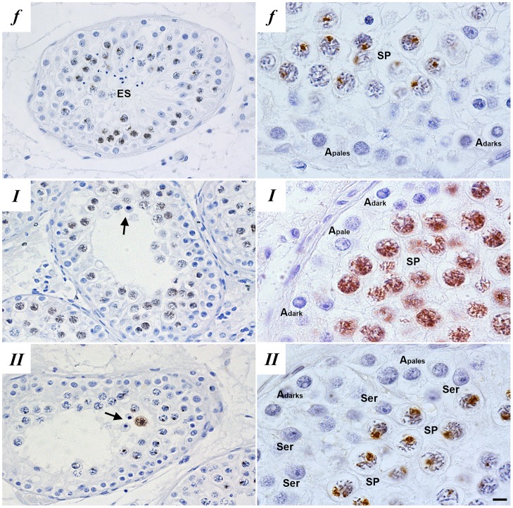
Fig. 1.
Histological evaluation of patient testis sections. Immunohistochemical localization of γH2AX in paraffin-embedded testis sections of fertile men (f) and patients with meiotic maturation arrest reveal two types of meiotic prophase arrest patients: type I (I) and type II (II) meiotic arrest. Type I meiotic arrest patients display meiotic prophase arrest and disturbed γH2AX distribution and no XY-body formation, type II meiotic arrest patients display meiotic prophase arrest but normal γH2AX distribution and XY-body formation. Left-hand panels show a global overview of the γH2AX staining in different germ cell populations within the testis and the right-hand panels show higher magnification images of γH2AX staining in spermatocytes. Depicted are: Sertoli cells (Ser), elongated spermatids (ES), Apale and Adark spermatogonia, spermatocytes (SP) and apoptotic spermatocytes (arrows). Scale bars: 3 μm (left); 10 μm (right).

