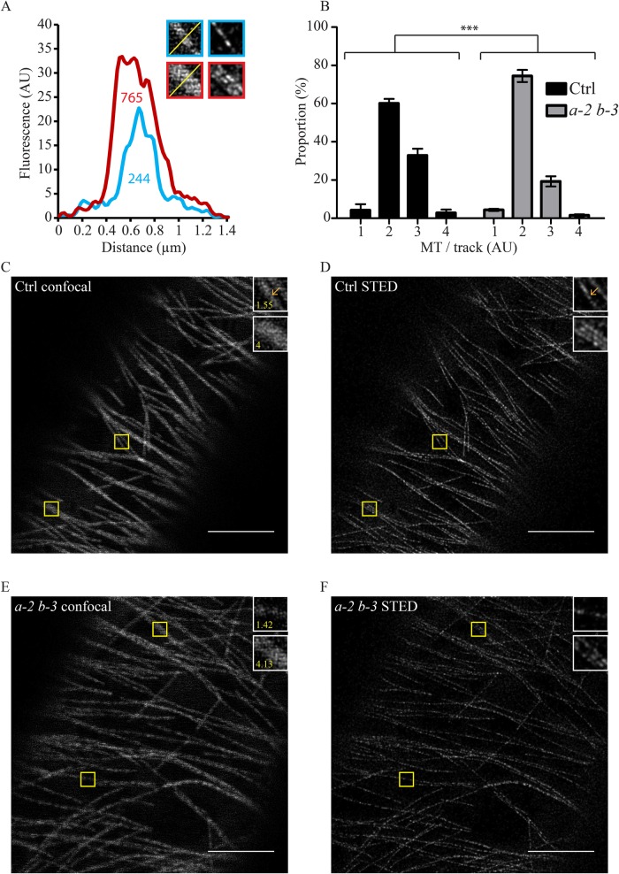
Fig. 2.
Bundling is impaired in eb1a-2 eb1b-3 double mutant. (A) The graph shows that the number of MTs within a fluorescence track is proportional to the area under the plot profile [values indicated under the curves in arbitrary unit (AU)]. STED imaging reveals that the red-bordered track is made of at least three parallel MTs, whereas the blue-bordered one is made of at least one MT, which is consistent with the plot profiles and the related values. (B) Frequency distribution of the number of MTs per track in both genotypes (n=270 for the control and n=276 for the double mutant) based on the proportionality demonstrated in A. Asterisks indicate statistically significant differences (P=0.0006, Chi-square), bars represent s.e.m. Pictures showing confocal images (C,E) and STED images (D,F) of the same cell in control (C,D) and eb1a-2 eb1b-3 double mutant (E,F) hypocotyl epidermal cells. Insets (yellow squares) show details of MT. Yellow number in insets indicate the number of MTs in the selection based on the method described in A. Scale bar: 5 µm.

