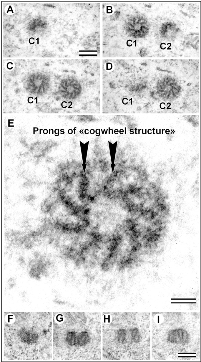
Fig. 2.
Fine centriole structure in the epithelial somatic cell of male A. calandrae larvae. (A–D) Four consecutive serial cross-sections of two centrioles, view from the distal ends of the both centrioles. (E) Cross-section of the centriole from panel C (C2) at high magnification. (F–I) Four consecutive serial sections parallel to the centriole axis. C1, centriole 1; C2, centriole 2. Scale bars: 200 nm for A–D,F–I. 50 nm for E.

