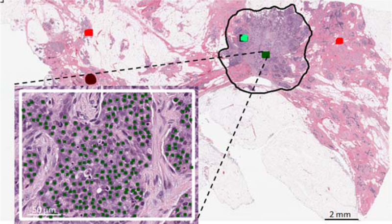
Figure 1.

Subset of nuclei annotations by expert pathologist. Centroids of different nuclei classes (malignant epithelial in this demonstration) were marked by the pathologist. Regions of interest were selected from within the tumor bed regions (black contour). [Color figure can be viewed at wileyonlinelibrary.com]
