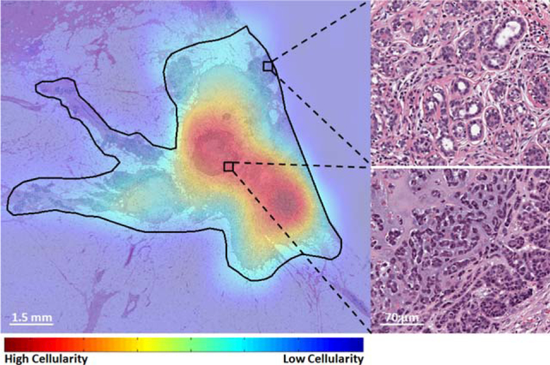
Figure 6.

Cellularity assessment heat map from within a tumor bed region (black contour) on a whole slide image with two representing benign (top-right) and malignant (bottom-right) regions. [Color figure can be viewed at wileyonlinelibrary.com]

Cellularity assessment heat map from within a tumor bed region (black contour) on a whole slide image with two representing benign (top-right) and malignant (bottom-right) regions. [Color figure can be viewed at wileyonlinelibrary.com]