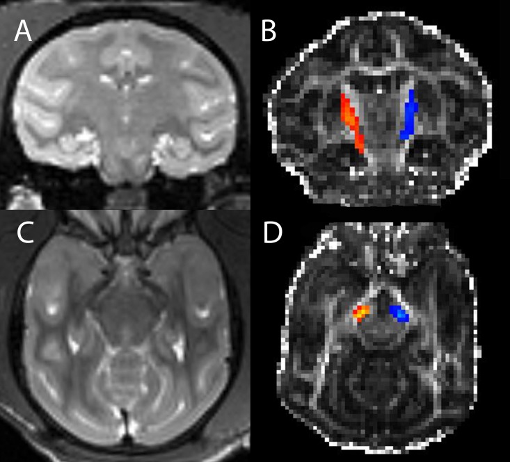
Fig 1. Nigrostriatal track volume.
T2 weighted coronal image through the striatum (A) and FA weighted image (B) with superposed nigrostriatal track volumes (right in orange and left in blue) in a single monkey. Analogous transverse images at a level above the substantia nigra are shown in (C) and (D).

