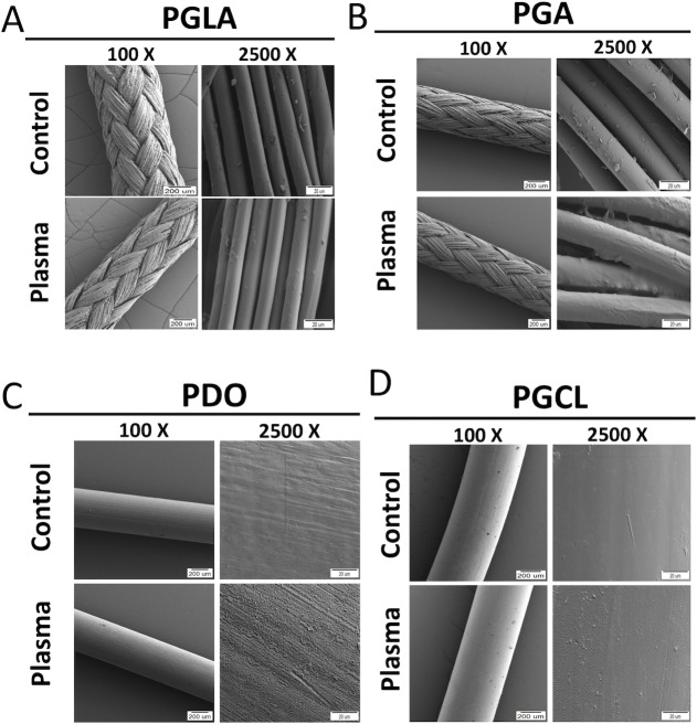
Fig 9. Scanning electron microscopy images of suture samples before and after NTAP treatment.
SEM images of (A) PGLA, (B) PGA, (C) PDO and (D) PGCL sutures were obtained before and after 7-minute NTAP treatment at 100X and 2500X magnifications to evaluate structural and surface characteristics, respectively.

