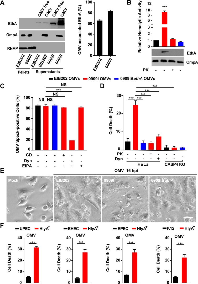
Fig 3. Hemolysin associates with OMVs to promote caspase-4 activation.
(A-B) Immunoblots for EthA in different fractions of E. tarda culture at 10 h post-inoculation (A) and assay for hemolytic activity and immunoblots for EthA in OMVs, treated with PK (5 μg/mL, 5 min) or not (B). (C) Immunostaining for intracellular OMVs using anti-OmpA antibody and quantification of OMV speck-positive cells in HeLa cells incubated with indicated E. tarda OMVs (20 μg, 1 × 10⁵ cells, 4 h) in the presence of CD (10 μM), EIPA (30 μM), Dyn (80 μM), or not. Percentage of cells containing OMV specks was calculated for at least 500 cells. (D and F) LDH release detected in wild-type or Caspase-4-/- HeLa cells incubated with purified E. tarda OMVs (50 μg, 1 × 10⁵ cells, 20 h) pretreated with PK (5 μg/mL, 5 min), Dyn (80 μM), or not (D), or purified E. coli OMVs (50 μg, 1 × 10⁵ cells, 20 h) (F). (E) Morphology of HeLa cells infected with indicated E. tarda OMVs (50 μg, 1 × 10⁵ cells, 16 h). Scale bar, 20 μm; arrows, pyroptotic cells. Graphs show the mean and s.e.m. of triplicate wells and are representative of three independent experiments. *p < 0.05, **p < 0.01, ***p < 0.001; NS, not significant (two-tailed t-test).

