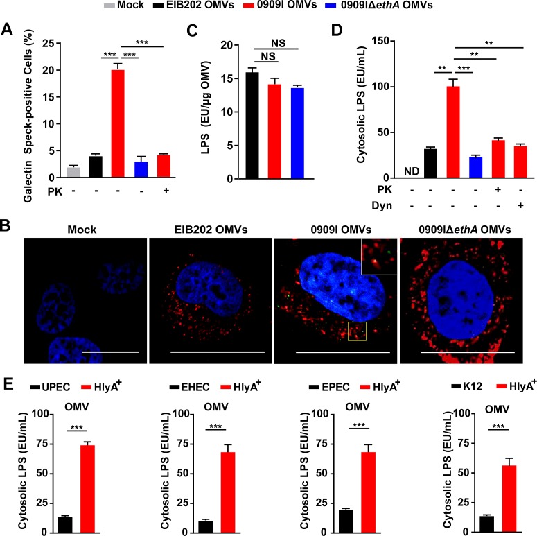
Fig 4. Hemolysin impairs OMVs-containing vacuole integrity for cytosolic access of LPS.
(A) Quantification of galectin speck-positive cells in HeLa cells expressing GFP-tagged gelectin-3, incubated with E. tarda OMVs (50 μg, 1 × 10⁵ cells, 20 h) treated by PK (5 μg/mL, 5 min) or not. Percentage of cells containing galectin specks was calculated for at least 500 cells. (B) Observation of intracellular galectin (green)-associated OMV specks using anti OmpA antibody (red). Scale bar, 20 μm; the white box indicates the co-localized signal between galectin and OMVs. (C) LPS quantification by LAL assay in purified E. tarda OMVs. (D-E) LPS quantification by LAL assay in the cytosol extracted by digitonin fractionation from Caspase-4-/- HeLa cells, incubated with E. tarda OMVs (50 μg, 1 × 10⁵ cells, 20 h) treated with PK (5 μg/mL, 5 min), Dyn (80 μM), or not (D), or E. coli OMVs (50 μg, 1 × 10⁵ cells, 20 h) (E). Graphs show the mean and s.e.m. of triplicate wells and are representative of three independent experiments. *p < 0.05, **p < 0.01, ***p < 0.001; NS, not significant (two-tailed t-test).

