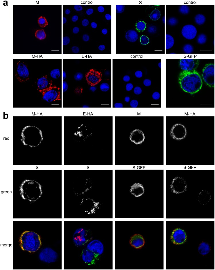
Fig 2. Expression and co-localization of HCoV-NL63 proteins in insect cells.
Confocal microscopy analysis of HF cells infected with single rBVs: M, S, M-HA, E-HA, and S-GFP (a) or co-infected with several rBVs. Single channels images and merged pictures are shown (b). M was detected with anti-M antibody (shown in red); M-HA and E-HA proteins were detected with anti-HA antibody (shown in red); S was detected with anti-S antibody and S-GFP (shown in green); nuclei are presented in blue. Scale bar: 10 μm.

