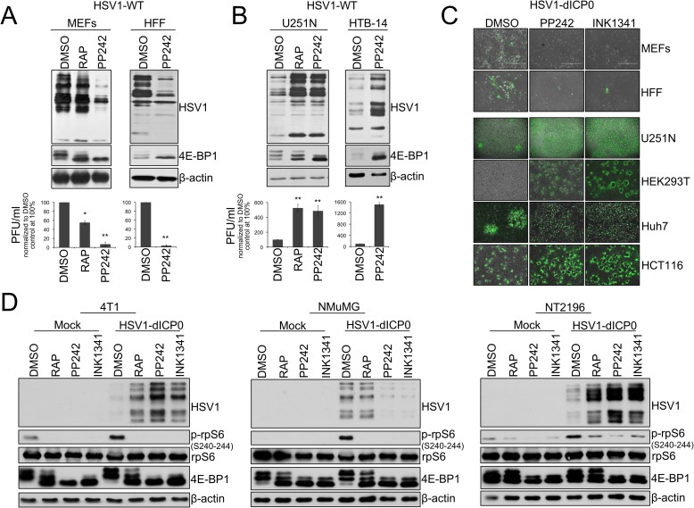
Fig 1. Active-site mTOR inhibitors (asTORi) augment HSV1 infection specifically in transformed cells.
(A) Primary mouse embryonic fibroblasts (MEFs), human foreskin fibroblasts (HFF), and (B) human glioblastoma cell lines U251N and HTB-14 were pretreated with DMSO, rapamycin (RAP 100nM), or the asTORi PP242 (2μM) for 30min followed by infection with wild type HSV1 at a MOI of 0.1 for 48 hours in presence of the inhibitors. Viral infection was monitored by Western blot using antibodies against HSV1 antigens (top panel - 4E-BP1 and β-actin expression were used to monitor drug efficacy and loading, respectively), and plaque titration (bottom panel—results are presented as titers normalized to DMSO control set at 100% ± SD (n = 3)). (C) Non-transformed MEFs and HFF, as well as different transformed human cell lines, were infected with GFP-expressing HSV1-dICP0 for 48 hours at a MOI of 0.1. Resulting infection was assessed by fluorescence microscopy. (D) Transformed (4T1 and NT2196) and non-transformed (NMuMG) mouse mammary cell lines were pretreated with DMSO, rapamycin (100nM), PP242 (2μM) or INK1341 (100nM) for 30 min followed by infection with a GFP-expressing HSV1-dICP0 (0.1 MOI for 48 hours in presence of the inhibitors). Viral protein synthesis was monitored by Western blot against HSV1. Drug efficacy was monitored by phosphorylation of rpS6 and 4E-BP1. Total rpS6 and β-actin expression were used as loading controls.

