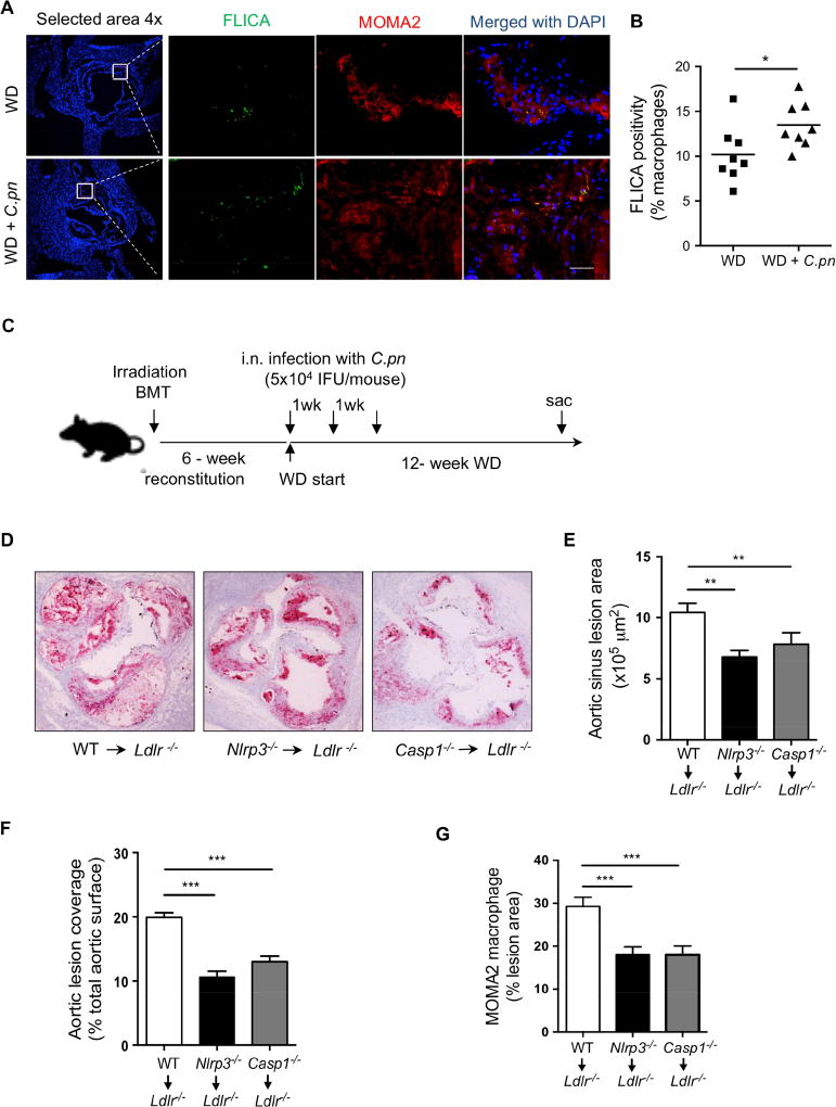
Figure 2. Nlrp3-deficiency in hematopoietic cells prevents atherosclerosis acceleration by C.pn infection.
(A) Caspase-1 activity was assessed by FLICA (green) and in macrophages (MOMA-2; red) in atherosclerotic lesions of Ldlr−/− mice fed a WD for 16 weeks with and without C.pn infection (n=6). Representative images for Caspase-1 positivity in lesion macrophages are shown. Scale bar = 50 μm.
(B) Quantification of active caspase-1+ cells in lesion macrophages.
(C) BM from WT, Nlrp3−/−, or Casp1−/− mice were transplanted to irradiated Ldlr−/− mice. 6-weeks after reconstitution, mice were put on WD (12-weeks). All groups were infected with intra-nasal C. pn (5 × 10⁴ IFU/mouse) weekly for a total of three times (beginning at the onset of WD). (n=11–12).
(D) Representative Oil Red O staining of aortic root plaques.
(E) Quantification of aortic root lesion area (n= 11 per group)
(F) Quantification of aortic en face lesion area (n=12 per group).
(G) Quantification of macrophage content in the aortic root lesions (n=10 per group).
All data are mean±SD. Significance was determined using Student’s t-test (B) or One-Way ANOVA with Tukey’s post-hoc test (E-G). *p<0.05, **p<0.01, ***p<0.001. Both male and female mice were used.

