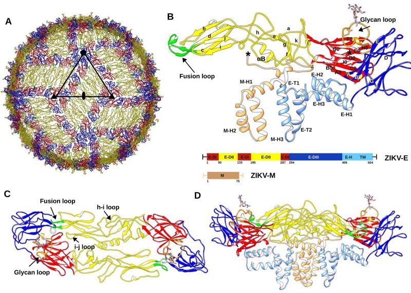
Figure 1. Overall Structure of ZIKV.
(A) ZIKV structure highlighting the herringbone pattern formed by 6 E–M heterodimers. The icosahedral asymmetric unit, is outlined by a black triangle. The structures of the E proteins are shown as Cα backbone traces. Domains E-DI, E-DII and E-DIII of each E protein are shown in red, yellow and blue, respectively. The fusion loop of each E protein molecule is shown in green.
(B) Secondary structural elements of one E–M heterodimer are labelled. Secondary structure elements on E-DI, E-DII and E-DIII are labelled A0–I0, a–l and A–G, respectively. The stem and transmembrane helices (E-H1, E-H2, E-H3, E-T1, E-T2) of the E protein and the M protein (M-H1, M-H2, M-H3) are colored in light blue and light brown, respectively. Residue numbering and domain definitions are shown as a linear peptide. Domains E-DI, E-DII and E-DIII are colored as in Figure 1A.
Panels (C) and (D) show the top and side views of the dimer, respectively. The glycan loop, fusion loop, h–I loop and i–j loop are labelled. (See also Table S1.)

