
Figure 1.
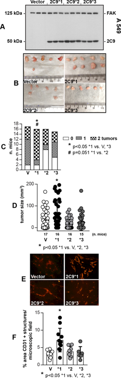
CYP2C9*1 promotes tumorigenesis. A, Cell lysates from A549 cells infected with empty adenovirus (Vector), or adenovirus carrying CYP2C9*1, CYP2C9*2, or CYP2C9*3 cDNAs were analyzed by Western blotting for expression of CYP2C9 and focal adhesion kinase (FAK, used as loading control) (n=3 infections). B, Images of tumors isolated from mice 14 days after receiving two s.c. injections of A549-Vector, −2C9*1, −2C9*2, or −2C9*3 cells. C, Tumor uptake was evaluated by counting the number of tumors in each mouse. The mice were divided into three groups: no tumors (0% uptake), one tumor (50% uptake), and two tumors (100% uptake). D, Tumor volume was evaluated with a caliper. Circles show individual tumors, while bars and errors show mean values and SD E, F, Tumor frozen sections were stained with anti-mouse CD31 antibody and vascularization was quantified as described in the Methods. Circles show individual mice, while bars and errors show mean values and SD.