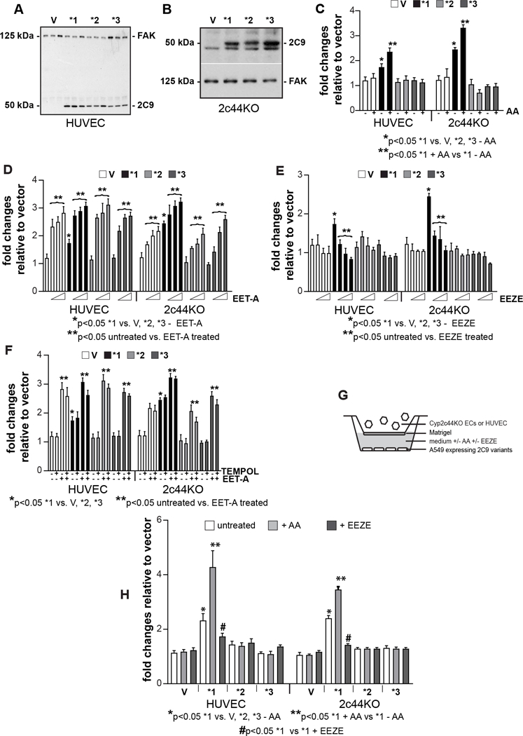
Figure 4.

Effects of CYP2C9* variants on EC proliferation and migration. A, B, Cell lysates from HUVEC (A) or Cyp2c44KO (B) ECs infected with empty adenovirus or adenovirus carrying CYP2C9*1, CYP2C9*2, or CYP2C9*3 cDNAs were analyzed by Western blotting for expression of CYP2C9 proteins and FAK. C-F, HUVEC or Cyp2c44KO ECs infected as described above were plated in 96-well plates in complete medium. After 12 hrs, the cells were incubated with serum-free medium with 3H-thymidine with or without AA (1 uM) (C), EET-A (1.25–5 μM) (D), 14,15-EEZE (1.25–5 μM) (E), or TEMPOL (4 μM) alone or in combination with EET-A (5 μM) (F). After 48 hrs, proliferation was determined as described (26). Values represent the mean ± SD of two experiments performed at least in triplicate. G, Schematic representation of the modified Boyden chamber assay used to evaluate migration of HUVEC or Cyp2c44KO ECs. The bottom well contains A549 cells infected with vector or the various CYP2C9* variants incubated with serum free medium that contained or not AA (1 uM) or 14,15-EEZE (5 uM). H, HUVEC or Cyp2c44KO ECs, added to the upper wells of Boyden chamber as described in G, were allowed to migrate for 4 hrs. The number of migrated cells was counted and expressed as the number of cells per microscopic field. Values are the mean ± SD of two experiments with at least eight microscopic fields evaluated.
