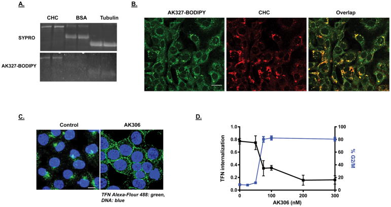
Figure 3.
A) AK3-BODIPY binding to CHC. Purified CHC, BSA and tubulin were run on duplicate SDS gels. The top gel was stained with SYPRO and the bottom gel was incubated with AK327-BODIPY (500 nM overnight in protein renaturation buffer). Both gels were viewed under UV/blue light illumination. B) Colocalization of CHC and AK3-BODIPY staining. YAMC cells were fixed and stained for CHC using Cy3 (red fluorescence). AK3-BODIPY was applied approximately 30 minutes prior to cell imaging. The BODIPY signal was found to concentrate in areas where CHC was highest. The bar shown is 20 μm. C) Endocytosis assay. HCT116 cells were treated with AK306 for thirty minutes, followed by addition of transferrin (TFN) conjugated to Alexa-488 (for thirty minutes). Cells were then fixed and analyzed by confocal microscopy. Bar indicates 20 μm. D) Dose response comparing the inhibition of TFN-Alexa 488 internalization and G2/M arrest.

