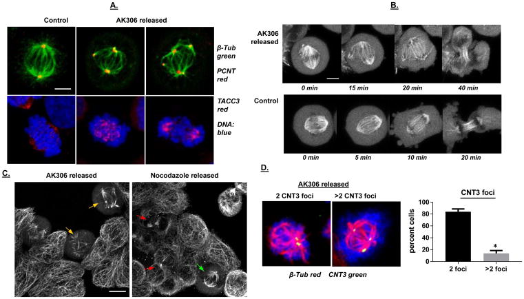
Figure 5.
A) Analysis of mitotic structures following release from AK306 arrest. Release from AK306 arrest causes ectopic localization of centrosome-related proteins and abnormal mitosis. Confocal images of representative cells were collected after immunostaining for pericentrin and TACC3. Mitotic abnormalities include multipolar spindles, an inability to line up chromosomes at the metaphase plate, and multipolar cell division. The bar shown is 5 μm. B) Time-lapse images showing tripolar mitosis after removal of AK306, compared to normal bipolar mitosis. Cells were transduced with CellLightTubulin-GFP construct to label microtubules. Cells were then treated with AK306 and released. The top row shows the formation of aberrant tripolar spindle and abnormal cytokinesis in an AK306 released cell. The bottom row shows normal mitotic progression in a non-treated cell. The bar indicates 5 μm. C) Impeded cell division after AK306 withdrawal in contrast to nocodazole withdrawal. Cells were treated with either AK306 or nocodazole and then released for two hours. Cells were fixed and stained with antibody to tubulin. Cells released from AK306 formed aberrant multipolar spindles (yellow arrows), whereas cells released from nocodazole were able to achieve metaphase (green arrow) and complete cytokinesis (as indicated by midbody formation; red arrows). The bar shown indicates 10 μm. D) Cell were released from AK306 arrest as in 5C, and then stained for tubulin (red) and centrin-3 (green). Cell with aberrant spindles were then scored for the number of centrin-3 foci. Representative images are shown in the left panels and the quantified data in the graph. Most of the cells showed aberrant spindles in the absence of centrin over-replication.

