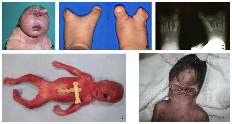
Figure 1.
A) Trisomy 13 fetus with cyclopia and proboscis. Reprinted by permission from Springer Nature (Capobianco et al., 2007). B) Bilateral split foot. Reprinted by permission from Oxford University Press (Hong et al., 2016). C) Bilateral split foot and the absence of second digit phalanges bilaterally on X-ray. Reprinted by permission from Oxford University Press (Hong et al., 2016). D) Frontal view of a 24-week-old fetus. Note cyclopia (synophthalmia), ambiguous genitalia, and partial syndactyly of 2nd and 3rd toes on the right Reprinted by permission from John Wiley and Sons (Weaver et al., 2010). E) Agnathia–otocephaly complex with cyclopia, agnathia, microstomia and ventromedial ear position. Reprinted by permission from BMJ Publishing Group Ltd. (Wai & Chandran, 2017).

