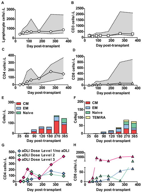
Figure 2. Reconstitution of immune cell subsets after T-cell depleted haploidentical HSCT and infusion of aDLI.
(A)–(D) Absolute numbers of lymphocytes (A), CD3+ (B), CD4+ (C) and CD8+ T-cells (D) in patient peripheral blood after T-cell depleted haploidentical HSCT and infusion of aDLI. Medians and interquartile ranges are shown for 16 patients eligible for immune reconstitution studies.
(E)–(F) Absolute numbers of CD4+ (E) and CD8+ (F) T-cells in patient peripheral blood after T-cell depleted haploidentical HSCT and infusion of aDLI with naive, central memory (CM), effector memory (EM) and T effector memory-RA (TEMRA) phenotypes. Medians are shown for 16 patients eligible for immune reconstitution studies.
(G)–(H) Absolute numbers of CD4+ (G) and CD8+ T-cells (H) in patient peripheral blood after T-cell depleted haploidentical HSCT and infusion of aDLI. Median values are shown for 16 patients eligible for immune reconstitution studies grouped according to aDLI dose.

