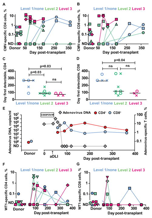
Figure 3. Reconstitution of virus- and tumor antigen-specific T-cells after T-cell depleted haploidentical HSCT and aDLI.
(A)–(B) Frequencies of CMV-specific IFN-γ+ CD4+ (A) and CD8+ (B) T-cells in donor and patient peripheral blood after T-cell depleted haploidentical HSCT and infusion of aDLI. Individual results are shown for patients who received transplants from CMV-seropositive donors and were eligible for immune reconstitution studies and one patient who received a transplant from a CMV-seronegative donor (P02030). Patients are colour coded according to the dose of aDLI. ND= not detectable.
(D–E) First time points when virus-specific IFN-γ+ T-cells were detectable in peripheral blood of individual patients after transplant. Crosses denote CMV-specific T-cells and circles denote VZV-specific T-cells. Results are shown for 13 patients with CMV- and/or VZV-seropositive donors who were eligible for immune reconstitution studies. Values are grouped and colour coded according to the dose of aDLI. Horizontal lines are medians.
(F–G) Frequencies of WT1-specific IFN-γ+ CD4+ (A) and CD8+ (B) T-cells in donor and patient peripheral blood after T-cell depleted haploidentical HSCT and infusion of aDLI. Individual results are shown for 14 patients eligible for immune reconstitution studies, colour coded according to the infused dose of viable donor CD3+ T-cells within aDLI/kg body weight.

