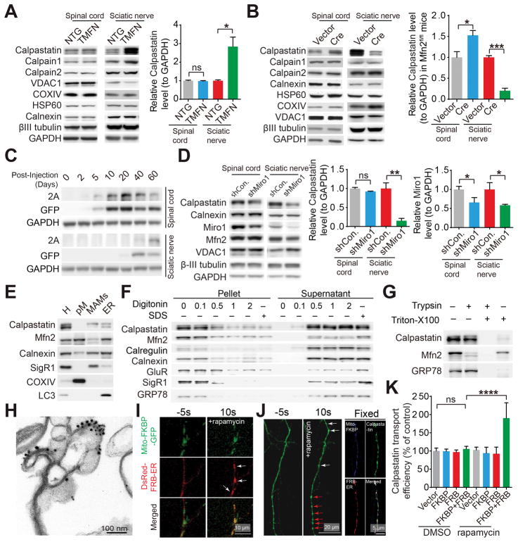
Figure 5. Mfn2 regulates the level of calpastatin in motor axons through the mitochondrial trafficking dependent active axonal transport.
(A and B) Representative immunoblot and quantification of calpastatin in endpoint G93A and dTg mice and age-matched NTG and TMFN mice (A) or 4–6 month old Mfn2fl/fl mice 12 days after injection with AAV1-GFP (Vector) or AAV1-Cre (Cre) (B). n = 3 or 4 mice per group. (C) Representative western blot detection of T2A tagged human calpastatin in lumbar spinal cords and sciatic nerves of 3–4 month old NTG mice at different days after AAV1-calpastatin injection. (D) Representative immunoblot and quantification of calpastatin in 3–4 month old NTG mice 12 days after injection of AAV1 encoding control shRNA (AAV1-shCon.) or AAV1-shMiro-1. n = 3 or 4 mice per group. (E–H) Representative western blot detection (E–G) and immunoelectron-microscopic analysis (H) of calpastatin (labeled by 15 nm gold particles) and Mfn2 (labeled by 10 nm gold particles) in MAMs isolated from pooled spinal cords of 4 NTG mice at 3–4 month old with or without indicated digitonin (w/v), triton X-100 (0.5% v/v) or trypsin treatment (F and G). H: total spinal cord homogenate; pM: highly purified mitochondria isolated from spinal cords. The purity of pM and MAMs was confirmed by EM (Figure S5A and S5B). (I and J) Representative confocal images of mitochondria and ER (I) or GFP tagged calpastatin in live cultured (I and J left panel) or fixed (J right panel) neurons 2 days after co-transfection with mito-FKBP-GFP and DsRed-FRB-ER (I, white arrows point ER recruited to mitochondria) or mito-FKBP-myc, Flag-FRB-ER and GFP tagged calpastatin (J, white arrows point newly formed calpastatin puncta while red arrows point enlarged pre-existing calpastatin puncta) before and after rapamycin treatment. (K) Quantification of calpastatin axonal transport (indexed by the relative ratio of the mean gray value of calpastatin in a segment of neuronal process 200 μm length beginning from the cell body of neurons to the mean gray value of calpastatin in the cell body of neurons) in neurons 2 days after transfection or co-transfection with mito-FKBP-myc, Flag-FRB-ER and GFP tagged with or without rapamycin treatment for 30 min. n = 15–24 neurons per group. Equal protein amounts of 20 μg were loaded for all blots. Data are means ± s.e.m., representative of triplicate experiments. Student’s t test or one-way analysis of variance (ANOVA) followed by Tukey’s multiple comparison test. *P < 0.05, **P < 0.01, ***P < 0.0001, ****P < 0.0001. ns, not significant.

