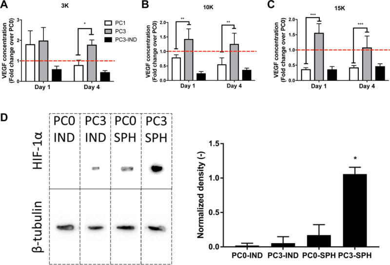
Figure 4. Proangiogenic potential of MSC spheroids is enhanced with hypoxic preconditioning.

VEGF concentration in conditioned media from gels cultured for 1 or 4 days containing preconditioned MSCs as (A) 3K cells/spheroid, (B) 10K cells/spheroid, and (C) 15K cells/spheroid; n=4. *p<0.05, **p<0.01, ***p<0.001. Data are normalized to VEGF concentrations of unconditioned PC0 spheroids, and red dotted line indicates PC0 group. (D) Western blot for HIF-1α expression. Bands are (left to right) individual MSCs maintained in ambient air (PC0 IND) or preconditioned for 3 days (PC3 IND), spheroids from unconditioned MSCs (PC0 SPH), and spheroids formed from hypoxic preconditioned MSCs (PC3 SPH). β-tubulin served as the loading control. Density of bands was determined using ImageJ and normalized to housekeeping band density for quantification of protein (right, n=3). *p<0.05 vs. all groups.
