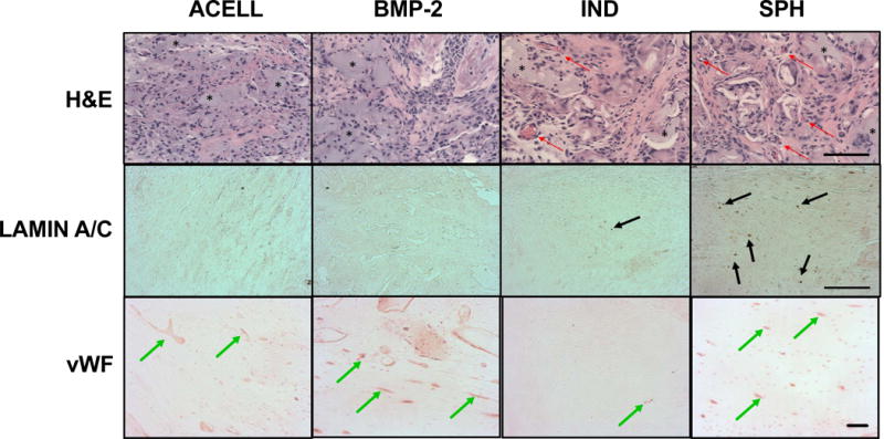
Figure 6. MSC spheroids stimulate vascularization and persist in vivo at 2 weeks in a critical sized femoral defect.

Representative H&E, Lamin A/C, and von Willebrand Factor (vWF) staining from acellular gels (ACELL), alginate gels containing BMP-2 (BMP-2), preconditioned individual MSCs (IND), or spheroids formed from preconditioned MSCs (SPH). Red arrows denote blood vessels, black arrows denote positive staining for human cells, and green arrows highlight positive staining for blood vessels. * indicates residual alginate. Images taken from the center of the defect. Scale bar = 100 μm for H&E; 200 μm for Lamin A/C and vWF.
