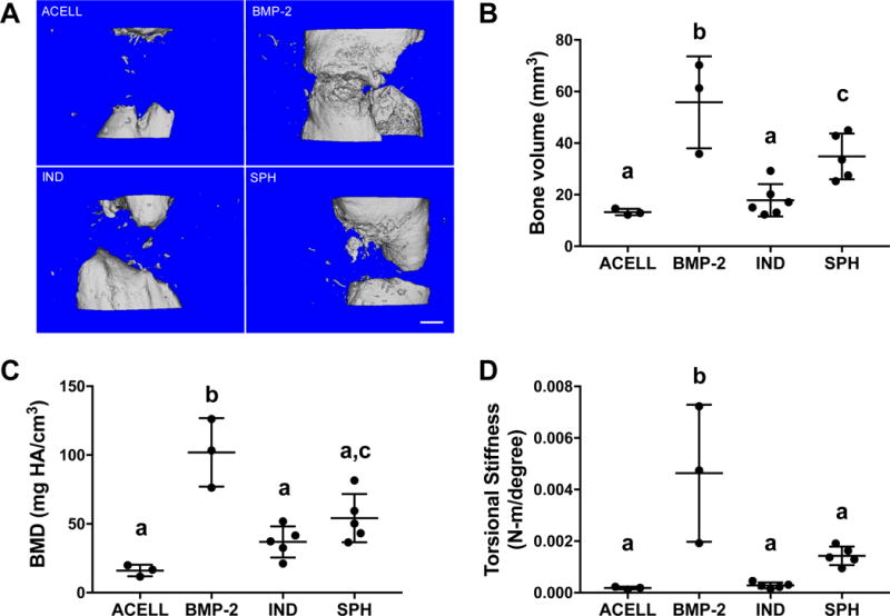
Figure 7. MSC spheroids formed from preconditioned cells promote bone formation in a critical-sized orthotopic bone defect.

(A) Microcomputed tomography images at week 12 in the defect space with quantified (B) bone volume and (C) bone mineral density. (D) Torsional stiffness of explanted femurs at 12 weeks; n=3-5 per group. IND = individual (non-aggregated) MSCs; SPH = spheroids. Significance is denoted by alphabetical letterings; groups with no significance are linked by the same letters, while groups with significance do not share the same letters.
