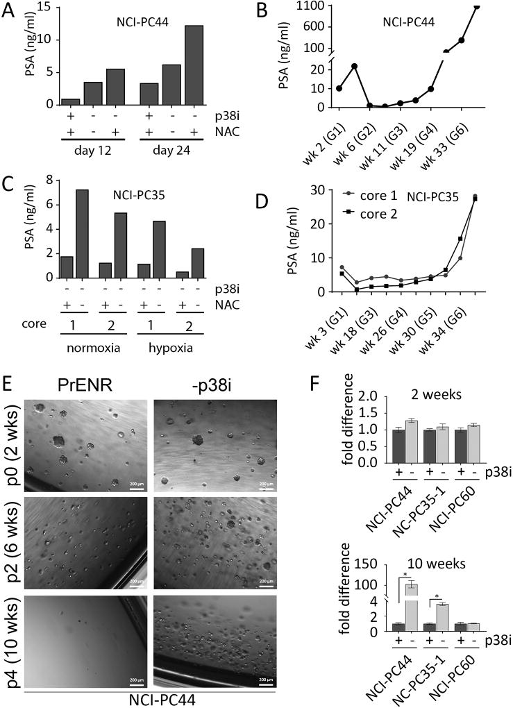
Figure 2.
Modified culture conditions improve growth of metastatic prostate cancer cells cultured directly from patient samples. A, NCI-PC44 spinal metastasis G1 cultured in PrENR (+p38i), −p38i, and −p38i −NAC conditions. PSA was measured at days 12 and 24 after initial collection and plating of the biopsy sample. B, Growth of patient derived organoids tracked by secreted PSA at several time-points, starting at G1 and continuing until G7. C, NCI-PC35 neck metastasis G1 grown in −p38i and −p38i −NAC in both normoxic (20% O2) and hypoxic (1% O2) atmospheres. The graph for NCI-PC35 shows data for two spatially separate biopsy cores (1 and 2), taken from the same tumor at the same time and maintained separately in culture. D, Growth of NCI-PC35 organoids tracked by PSA as described for (B). E, Patient-derived organoids NCI-PC44 cultured in PrENR (+p38i), or −p38i. Organoids were passaged every two weeks for ten weeks. The passage numbers in this figure indicate the number of passages from the initiation of this experiment and are not an indication of the number of passages for the organoid line overall. Representative bright field images were taken every two weeks prior to each passage. F, Patient-derived organoids NCI-PC44, NCI-PC35-1, and NCI-PC60 were cultured in the optimal media condition determined for each, or the optimal media +p38i. Organoids were passaged every two weeks for ten weeks, and quantified by CellTiter Glo 3D at two and ten weeks. Results are shown as fold difference between the +p38i and −p38i conditions. Error bars represent the standard error of the mean of three replicates. Student’s t-tests were performed; *P < 0.004

