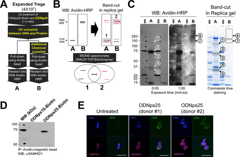
Figure 2. SAMHD1 is a cytoplasmic ODNps25 receptor to stabilize co-expression of Foxp3 and Helios in human Tregs.

(A) Two-step cross-link strategy to maximize pull-down efficacy of ODNps25-Biotin-interacting proteins. To crosslink ODNps25-biotin and its interacting protein, cells were exposed by UV (150 mJ/cm2), then incubated with disuccinimidyl suberate (DSS, 5 mM) for 30 min at room temperature. ‘A’ and ‘B' represent avidin-dependent pull-down proteins from single UV-crosslinked Tregs and two step cross linked Tregs by UV and DSS, respectively. (B) The principle to Identify proteins crosslinked with ODNps25-biotin covalently by comparing two different pull-down samples (A and B in Fig. 2a). Briefly, the gel positions of ODNps25-biotin and protein covalent complex would be determined by western blot of A and B samples with avidin-HRP, and the protein bands in sliced gel bands from replica Coomassie blue stained gel will be identified by MS/MS spectrometry. Overlapped protein IDs from the list of A and B band-cuts (1 and 2) would be considered as an ODNps25 receptor candidates, and the rest IDs in the lists will be excluded as a non-specific gel slice contaminants. Black thin line and red thin line represent gel contaminant with low molecular weight and with high molecular weight, respectively. (C) Western blot of A and B pull-down samples with avidin-HRP (left two membranes) and band-cut from replica SDS-PAGE gel (right Coomassie blue-stained gel). The numbers in the circle indicate specifically detected bands in A samples (1–5) and B samples (6 and 7) after blotting with avidin-HRP (1:10,000). The closed squares indicate the gel-cut positions corresponding to western bands in replica gel. pull-down samples were prepared as same as in Fig.2C. (D) Confirmation of SAMHD1 binding to ODNps25-biotin using avidin-dependent pull-down and western blot with anti-hSAMHD1 antibody (1:2,000). Data shown is one of two independent experiments with different donors. (E) Confocal microscopy of endogenous SAMHD1 (red) in ODNps25-FITC (green) treated expanded Tregs. Nuclei were counterstained with DAPI (blue). Scale bars, 10 µm. Data shown is one of three independent experiments with different donors.
