
Figure 4. SAMHD1 specifically binds to 3’-UTR of Foxp3 and Helios, and stabilize their post-transcription by TCR stimulation as well as ODNps25 addition.
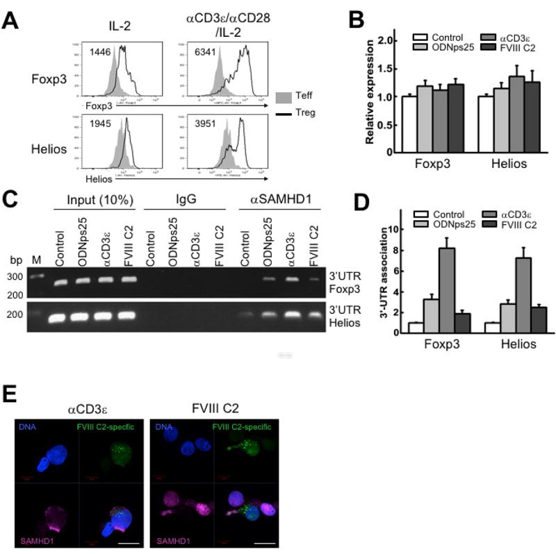
(a) Relative induction of Foxp3 and Helios proteins by in vitro CD3/CD28-stimulation in Tregs. Rested polyclonal Tregs, expanded as in Methods, were re-stimulated by adding anti-CD3ε (0.5 µg/ml), CD28 antibodies (0.2 µg/ml) and recombinant IL-2 (200 IU/ml) together with -irradiated PBMCs for 36 hrs. The number positioned in upper left corner in the histogram plots indicates median of MFI of Foxp3 or Helios, respectively. The plots shown is one of three independent experiments with different donors. (B) Relative expression of total mRNA of Foxp3 and Helios in 17195TCR-transduced Tregs stimulated with ODNps25, anti-CD3ε antibody, or FVIII C2 peptide. Preparation and in vitro expansion of 17195 TCR-transduced Tregs (17195 Tregs) is followed as shown in Rf. Expanded 17195 Tregs were rested in IL-2 deprived culture media overnight, and re-stimulated with ODNps25 (2 µM), anti-CD3ε antibody (0.5 µg/ml), or FVIII C2 peptide (1 µg/ml) for 36 hrs. Quantitative RT-PCR (qRT-PCR) of Foxp3 and Helios was performed with primers specific to 5’encoding region of Foxp3 and Helios (P1 and P2 for Foxp3 shown in Supplemental Fig.1). Data was normalized with external HPRT control. (C) RNA immunoprecipitation (RIP)-qPCR against SAMHD1 to detect 3’UTR of Foxp3 and Helios. RIP analysis was performed from the whole extract of re-stimulated 17195Tregs in (A). Amplification was performed by qPCR with primer specific to 3’UTR of Foxp3 and Helios (P3 and P4 for Foxp3 shown in Supplemental Fig.1). The experiments of B, C, and D are one of two independent experiments with different donors. (D) Relative quantification was measured by SYBR green-qPCR. (E) Confocal microscopy of SAMHD1 (red) in re-stimulated 17195 TCR transduced (green) Tregs. Cells were re-stimulated by adding anti-CD3ε antibody (0.5 µg/ml) or FVIII C2 peptide (1 µg/ml) for 36 hrs. FVIII-specific TCR expressed cells are recognized by the expression of green fluorescent protein (GFP). Nuclei were counterstained with DAPI (blue). Scale bars, 10 µm.