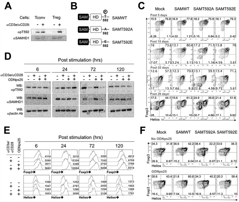
Figure 5. Thr592 phosphorylation of SAMHD1 due to TCR stimulation stabilizes the expression of Foxp3 and Helios in Tregs.

(A) The592 Phosphorylation of SAMHD1 by TCR stimulation. 2 wk-expanded conventional T cells (Tconv) or Tregs were re-stimulated with plate-coated anti-CD3ε /CD28 antibodies and IL-2 or IL-2 alone condition for 24 hrs. Western blots were performed with total proteins extracted in RIPA. Data shown is one of three independent experiments with different donors. (B) Expression constructs of phospho-deficient (SAMT592A) and phosphomimetic (SAMT592E) SAMHD1. (C) Stabilization of Foxp3 and Helios expressions in SAMWT, SAMT592A, or SAMT592E Tregs. The protocols for Transduction, expansion and FACS analysis is same as in Fig 3H. The day displayed above panels indicates post-time after primary stimulation. The plots shown is one of three independent experiments with different donors. (D) Kinetics of Thr592 phosphorylation of SAMHD1 in Tregs during CD3 stimulation with ODNps25. 2 wk-expanded Tregs (1×10⁶) were restimulated with indicated stimuli for given times. CD3 stimulation was applied by incubating the cells in plate-coated anti-CD3ε Ab. After incubation, the cells were harvested, and transferred to two tubes in an equal cell number. One was used to total protein extraction for the western blot, and the cells in the other tubes were stained for intracellular FACS of the Foxp3 and Helios. The number in histogram plots (numbers in the plot) represents mean fluorescence intensity (MFI) of histogram (E). (F) Stabilization of Foxp3 and Helios expressions in GFP+ SAMWT, SAMT592A, and SAMT592E Tregs in the addition of ODNps25 or not. The protocols for Transduction, expansion and FACS analysis is same as in Fig 3H except for the ODNps25 treatment (2 µM). Intracellular Foxp3 and Helios is measured at day 20 after initial stimulation. Data shown is one of two experiments with different donors.
