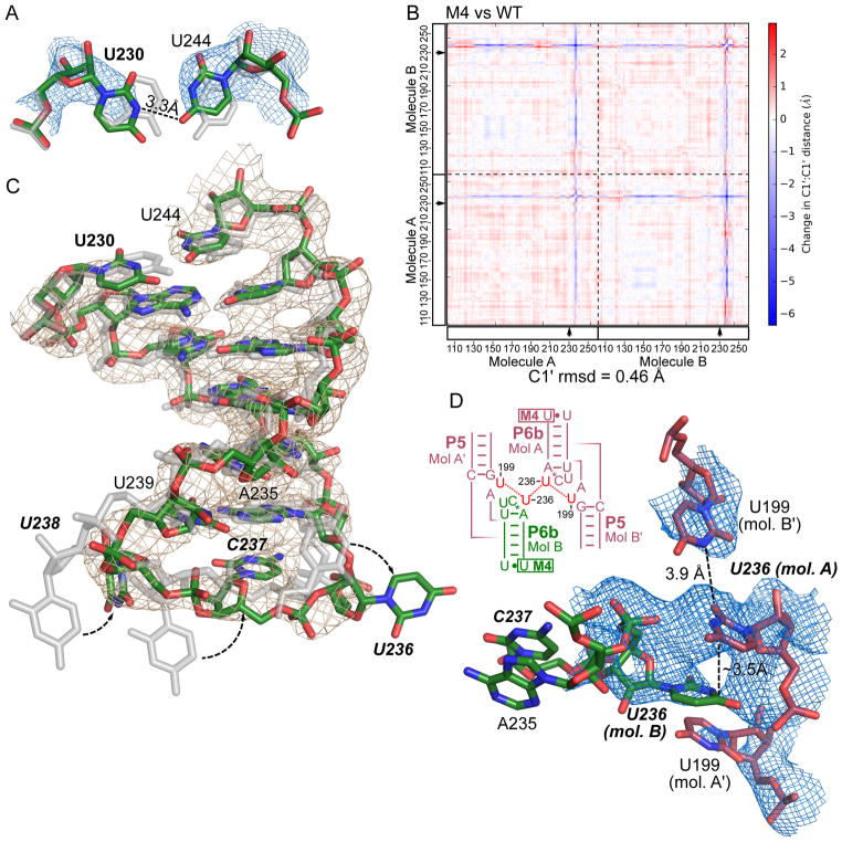
Figure 5. M4 weakens the P6b helix, allowing the L6b loop to establish new lattice contacts.
(A) The A230U mutation site in the 2.8-Å-resolution M4 structure (with atoms colored red, green and blue), compared to the WT structure shown in silver. The structures were superimposed by aligning all molecule B atoms outside of residues 230–244. Blue mesh shows the immediately surrounding 2Fo-Fc map for M4 contoured at 1.2 σ level. (B) Difference distance matrix plot between M4 and WT structures. Both molecules A and B in the asymmetric unit are shown, separated by dashed lines. Mutated sites are marked with arrows. Calculated r.m.s. deviation for all C1′ atoms in the asymmetric unit is shown below the plot. (C) The P6b stem loops in M4 and WT structures superimposed and colored as in (A). 2Fo-Fc map contoured at 1.2 σ level is shown as gray mesh. (D) New crystal contacts established by M4. Blue mesh shows 2Fo-Fc map contoured at 0.6 σ. A schematic of the new lattice contacts involving U236 in L6b and U199 in J5a/5 from four different molecules.

