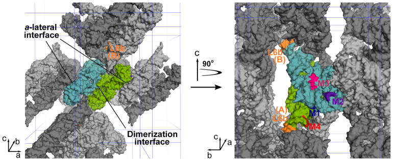
Figure 6. The new lattice contacts in crystals.
The left panel is a space-filling model of the WT P4–P6 molecules viewed along the b axis. The two unique molecules, A and B, are colored green and cyan, respectively. These two molecules effectively form a dimer via an extensive interface. Symmetry-related A and B molecules are indicated in dark and light gray, respectively. The right panel is a view of the RNA molecules in the left panel with a 90° rotation along the c axis. The P4–P6 dimer on the left side of the left panel is removed to illustrate the location of the contacts introduced by M2 (purple) and M3 (magenta, represented by residues 185–187) at the a-lateral interface on molecule B. Also visible in the right panel is the M1 (blue) mutation that establishes new contacts at the dimerization interface. The M4 (red) mutation allows the L6b loops (orange) in both molecules A and B to establish a new contact that involves four P4–P6 dimers (see also Figure 5D).

