Figure 1.
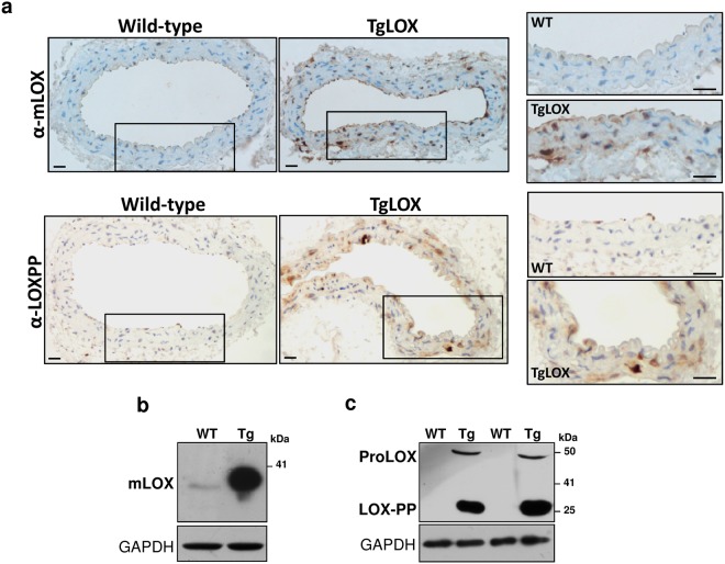
Mature LOX and LOX-PP were over-expressed in the vascular wall and in VSMC from TgLOX mice. (a) Representative immunohistochemical analysis showing LOX and LOX-PP staining (brown color) in aorta from both wild-type and TgLOX mice. The indicated areas are shown at high magnification (bars = 20 µm). (b,c) Mature LOX and LOX-PP protein levels were determined by Western-blot in VSMC supernatants from these animals. The position of the pro-enzyme (ProLOX), mature LOX (mLOX) and LOX-PP forms are indicated. GAPDH was analyzed as loading control. Representative immunoblots from 3 independent experiments were shown. WT: wild-type; Tg: TgLOX. Displayed blots are not cropped from different gels or different parts of the same gel and images conform the digital image and integrity policies of the journal.

