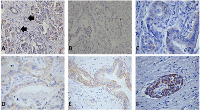
Fig. 5. Location and expression of PHB in normal and cancerous pancreatic tissues.
Immunohistochemistry was performed with PHB (1:5000) anti-rabbit antibodies. (magnitude ×200) a Localisation of PHB in normal pancreatic tissues, acinar cells and some ductal cells showed very weak cytoplasmic staining. b Very weak positive staining of PHB in cancer cells. c Weak expression of PHB shown by cytoplasmic staining. d Mild expression of PHB shown by plasma membrane and cytoplasm of cancer cells. e Strong expression of PHB shown by plasma membrane and cytoplasm of cancer cells. f PHB was also strong expressed in pancreatic islet cells

