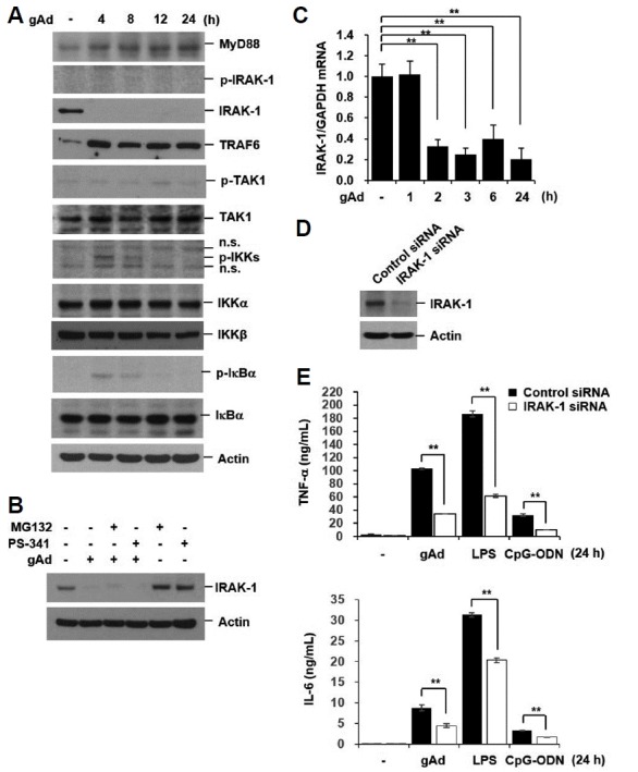
Fig. 4. gAd downregulates IRAK-1 mRNA and protein expression.

(A) RAW 264.7 cells were incubated with gAd (5 μg/ml) for the indicated times. (B) Cells were pretreated with MG132 (10 μM) and PS-341 (200 nM) for 1 h, and then stimulated with gAd (5 μg/ml) for 4 h in the presence or absence of MG132 and PS-341. Total cellular extracts were subjected to Western blot analysis for MyD88, p-IRAK-1, IRAK-1, TRAF6, p-TAK1, TAK1, p-IKKα/β, IKKα, IKKβ, p-IκBα, IκBα, and actin. (C) Cells were treated with gAd (5 μg/ml) for the indicated times. Quantitative real-time PCR for IRAK-1 and GAPDH was performed. Data represent the mean ± SD of triplicates. **P < 0.05 (D-E) RAW 264.7 cells were transiently transfected with control or IRAK-1 siRNAs. At 48 h after transfection, cells were treated with gAd (5 μg/ml), LPS (1 μg/ml), or CpG-ODN (1 μM) for 24 h. Total cellular extracts were subjected to Western blot analysis for IRAK-1 and Actin. TNF-α and IL-6 concentrations in culture medium were measured by multiplex bead assay. Data represent the mean ± SD of triplicates. **P < 0.05 Results are representative of three separate experiments.
