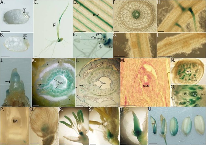
Fig. 2. Histochemical analysis of OsWOX13p::GUS reporter gene expression in plant development.

(A) Mature and (B) 2-day imbibed seeds. (C) A 5-day seedling. (D, E) Magnificent of a leaf blade (D) and longitudinal cut through a seed and stem (E) of a 5-day seedling. The white arrow in (E) indicates the position where GUS expression stopped on the crown root. (F) Cross-section of a crown root from a 7-day seedling. (G) Lateral root initiation in a 7-day seedling crown root. (H, I) Junctions with developed (H) and developing (I) lateral root in a 7-day seedling radicle. (J) Six-week-old stem with all developed leaves removed. The arrow shows the position of an auxiliary tiller where it is cross cut (K) and 12-μm-sectioned (L). The section in (L) was stained with toluidine blue. (M) The 12-μm cross-section of the SAM sample was stained with toluidine blue. (N) Hand section of a node from an unelongated stem. (O) Hand section of node Nth counted upward along an elongated stem. In node Nth, DVs only surround and connect to enlarged large VBs of leaf Nth at NVA. VBs of leaf (N+1)th are not enlarged and only pass through the node. (P) Panicles at the very early stage of inflorescent formation. (Q to T) Floret organs at stage (Q) 1 cm, (R) 12 cm, and (S) before and (T) after pollination. (U, left to right) Seeds at 2, 4, 6, 8, and 10DAP. Scale bar = 0.2 cm (a, b, c, e, j, t); 50 μm (F, H) or 200 μm (others). sc: scutellum, pl: primary leaf, ra: radicle, cr: crown root, me: mesocotyl, mv: main vien, NVA: nodal vascular anatomoses, VBs: vascular bundles, PLN and XLN: phloem and xylem of enlarged large VBs of leaf Nth, SN: enlarged small VBs of leaf Nth, L/SN+1: large/small VBs of leaf (N+1)th, DVs: diffuse vascular bundle, SAM: shoot apical meristem, IM: inflorescence meristem, DAP: day-after-pollination.
