Fig. 1.
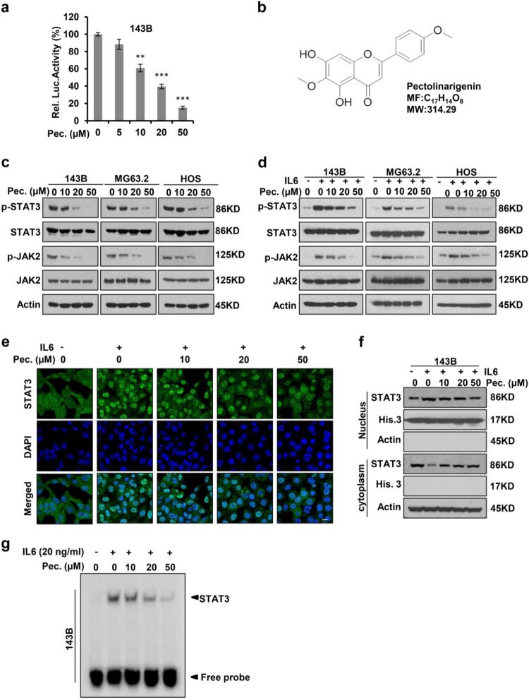
Pectolinarigenin inhibits STAT3 activity in osteosarcoma. a 143B cells were transfected with STAT3 Luciferase reporter gene plasmid and treated with different concentrations of pectolinarigenin for 24 h. The results were normalized to the Renilla luciferase activity (**P < 0.01; ***P < 0.001). b Chemical structure of pectolinarigenin. c A panel of osteosarcoma cell lines was exposed to the indicated concentrations of pectolinarigenin for 24 h. Cells were then lysed and applied to immunoblotting with the indicated antibodies. Actin was used as an internal control. d Osteosarcoma cell lines were pretreated with the indicated concentrations of pectolinarigenin for 24 h and then stimulated with IL6 (20 ng/μL) for 30 min. Whole cell extracts were prepared and subjected to western blot using the indicated antibodies. e 143B cells were seeded on gelatin-coated coverslips and pretreated with pectolinarigenin for 24 h followed by stimulating with IL6 (20 ng/μL) for 30 min. The coverslips were examined by a confocal microscopy. Anti-STAT3 antibody (green) was used to locate endogenous STAT3. Cell nuclei were stained with 4′,6-diamidino-2-phenylindole (DAPI). Scale bar, 20 μm. f 143B cells were treated with pectolinarigenin for 24 h, and the cytoplasmic and nuclear extractions were subjected to immunoblotting to detect the level of STAT3. g 143B cells were pretreated with pectolinarigenin and stimulated with IL6. An EMSA assay was performed to analyze STAT3 DNA-binding activity

