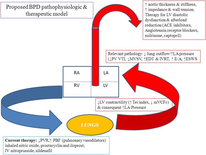
Figure 1.

BPD pathophysiologic and therapeutic model (PVR – pulmonary vascular resistance, PBF – pulmonary blood flow, LV – left ventricle, LA – left atria, RV – right ventricle, RA – right atria, PV – pulmonary vein, VTI – velocity time integral, MVSV – mitral valve stroke volume, EDT – E wave deceleration time, IVRT‐isovolumic relaxation time, ESWS – end systolic wall stress, mVCFc – mean velocity of circumferential fiber shortening, ACE – angiotensin‐converting enzyme). With permission from SAGE journals. Sehgal et al. A new look at Bronchopulmonary dysplasia: Postcapillary pathophysiology and cardiac dysfunction. Pulm. Circ. 2016; 6:508–15.
